Pain relief can start here.
A Holistic approach.
It serves to remember that we, as therapists, do not heal anything. We simply recognize, respect, and support the body's 'built-in' healing ability.
Corrective Massage recognizes the importance of balanced relationships, movements, and interactions throughout the body's soft tissues*. These relationships include natural anatomical positions and functionally balanced movement unimpeded by soft tissue tension.
Functional balance implies that our soft tissues are working harmoniously to support pain-free, healthy movement and function.
In Taum's world of Corrective Massage, considering all bodily movement as an orchestration of soft tissue relationships is a fundamental principle and starting point.
These foundational principles include familiarity with Equilibrium and Homeostasis. AKA Balance.
For example: Walking initially appears to be a simple activity. It actually involves a complex orchestration of numerous soft tissues.
A symphony of movement:
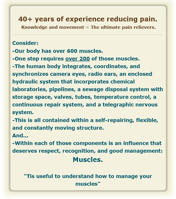
Our body requires over 200 of our 600 muscles to take one step. Of those 200, many serve as compensating adapters working in the background to keep us upright as we walk. While all our weight is on the right foot, those background muscles are adapting and counterbalancing so we do not fall over.
Each and every soft tissue plays a vital role in the body's ability to move and adapt to unbalanced tensions.
Our bodies can adapt to those tensions.
But only so far...
Limping is an example of crossing the adaptation line.
Pain serves as an alert that unbalanced tension has exceeded the body's ability to adapt, and corrective therapy is required.
The foundational focus of this unique therapy is to interpret the alert and then identify and correct the soft tissue tensions and imbalances that have created those alerts.
Corrective massage has repeatedly proven to be an efficient and effective method to reduce and relieve pain.
I hope this information serves you and those you serve.

*Muscles, tendons, ligaments, membranes, and viscera.
Pain relief can start here.
A Holistic approach.
It serves to remember that we, as therapists, do not heal anything. We simply recognize, respect, and support the body's 'built-in' healing ability.
Corrective Massage recognizes the importance of balanced relationships, movements, and interactions throughout the body's soft tissues*. These relationships include natural anatomical positions and functionally balanced movement unimpeded by soft tissue tension.
Functional balance implies that our soft tissues are working harmoniously to support pain-free, healthy movement and function.
In Taum's world of Corrective Massage, considering all bodily movement as an orchestration of soft tissue relationships is a fundamental principle and starting point.
These foundational principles include familiarity with Equilibrium and Homeostasis. AKA Balance.
For example: Walking initially appears to be a simple activity. It actually involves a complex orchestration of numerous soft tissues.
A symphony of movement:
Our body requires over 200 of our 600 muscles to take one step. Of those 200, many serve as compensating adapters working in the background to keep us upright as we walk. While all our weight is on the right foot, those background muscles are adapting and counterbalancing so we do not fall over.
Each and every soft tissue plays a vital role in the body's ability to move and adapt to unbalanced tensions.
Our bodies can adapt to those tensions.
But only so far...
Limping is an example of crossing the adaptation line.
Pain serves as an alert that unbalanced tension has exceeded the body's ability to adapt, and corrective therapy is required.
The foundational focus of this unique therapy is to interpret the alert and then identify and correct the soft tissue tensions and imbalances that have created those alerts.
Corrective massage has repeatedly proven to be an efficient and effective method to reduce and relieve pain.
I hope this information serves you and those you serve.
*Muscles, tendons, ligaments, membranes, and viscera.
|
Taum Sayers, CMT |
|
|
Site Overview
Appointments
Any improvement in balance can reap big rewards.
The sessions last approximately 30-45 minutes.
More Information
FAQ's
Some Frequently asked questions.
"How often should I stretch?"
"How do I know if I am doing..."
More
Self Help
With our present situation and
the challenges to our immune system,
this remedial core movement...
More Information
Covid-19
The information surrounding the
Covid-19 virus continues to
evolve and confuse...
Continue
Classes
For Massage Therapists, actually anyone interested in pain management.
Click here for information on Taum's classes.
Resources
Taum Sayers and others that you might find helpful in your pusuit of healthy balance...
Continue reading